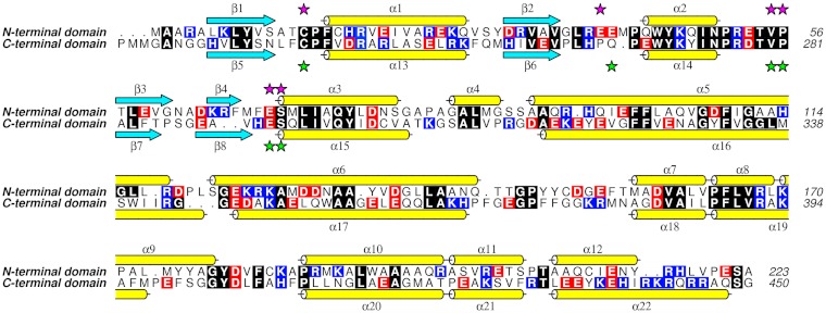
Fig. 2.
Sequence alignment of TDR1 domains I and II. Cyan arrows indicate β-strands and yellow cylinders α-helices. Selected residues involved in binding glutathione are indicated by purple stars, for domain I, and green stars, for domain II. Strictly conserved residues are encased in black, acidic residues in red and basic in blue. The alignment was performed using MUSCLE (28) and the figure produced with ALINE (30).

