Fig. 2.
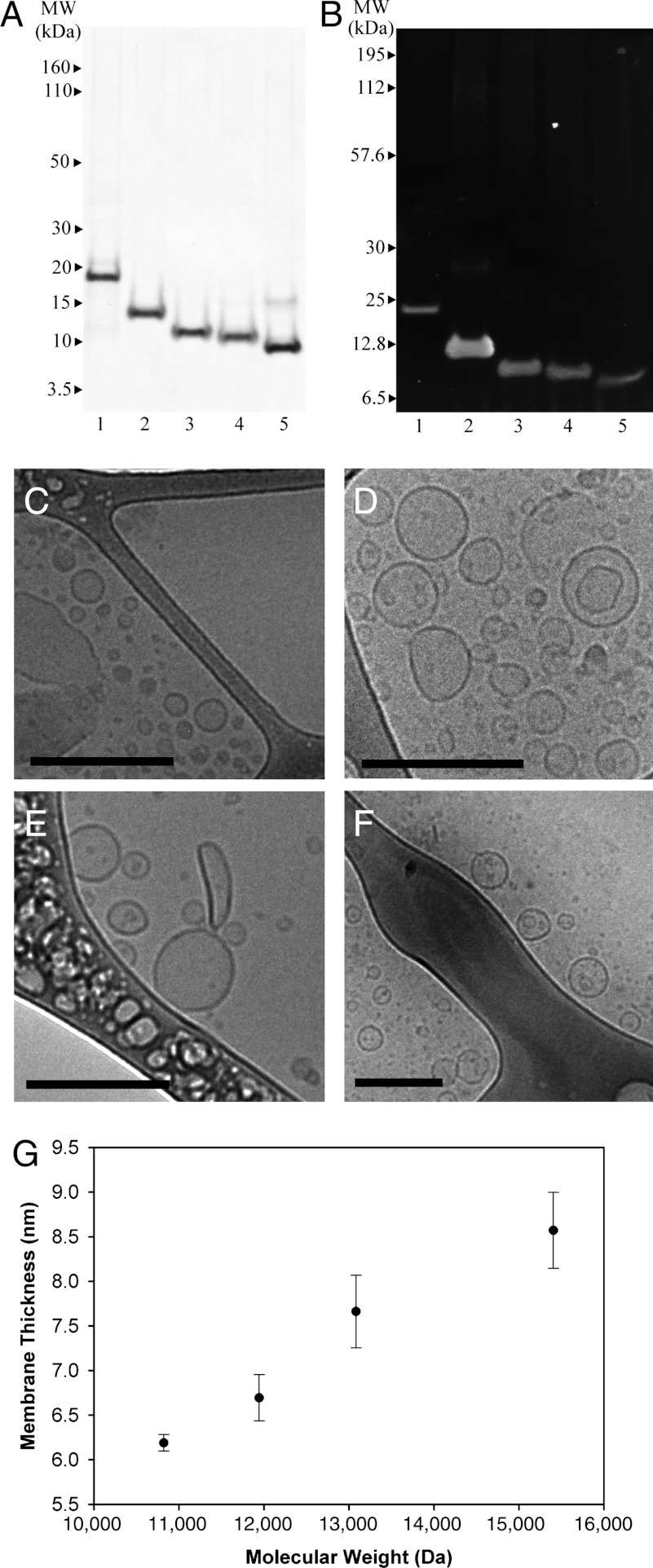
Protein identification and cryo-TEM evidence of self-assembled protein nanovesicles. (A) Protein purity was measured after organic solvent purification to be > 95% with SDS/PAGE. (B) Oleosin mutant identity was confirmed through Western blotting using an anti-6-His antibody. The lanes for both gels are (1) 42-87-57 (wild-type oleosin), (2) 43-65-33, (3) 33-65-23, (4) 28-65-18, (5) 23-65-13. Cryo-TEM micrographs indicate vesicle formation across the entire P = 65 family. All scale bars are 200 nm. (C) 23-65-13 and (D) 28-65-18 coexist as sheets (Fig. S3) and vesicles in PBS (140 mM) after injection. (E) 33-65-23 and (F) 43-65-13 form vesicles in PBS (140 mM). (G) Increasing the molecular weight of protein at constant hydrophobic block length increases the hydrophilic block fraction and leads to thicker vesicle membranes as measured through cryo-TEM. Protein hydrophilic fraction: 23-65-13: 35.4%, 28-65-18: 41.4%, 33-65-23: 46.3%, and 43-65-33: 53.9%. Error bars represent one standard deviation (N = 6).

