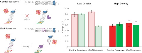
Fig. 5.
Electrocatalytic detection of sequence-specific restriction enzyme RsaI activity. Left: Sequences for the control and RsaI binding sequence are indicated. The binding site for RsaI is indicated in purple. All electrodes were incubated with BSA (1 mM for 1 h to minimize any signal attenuation observed from nonspecific protein binding). Electrodes were either Left untreated (red) or treated (green) with RsaI (20 μL of 50 nM) for 2 h. Right: The accumulated charge from the chronocoulometry data (V = -450 mV for 10 s) of MB-DNA in the presence of 25 μM hemoglobin for the control sequence is plotted for both high-density (dark) and low-density (light) DNA films. High-density films were formed by the addition of 100 mM MgCl2 during DNA film self-assembly. The analogous data for the RsaI sequence is also presented.

