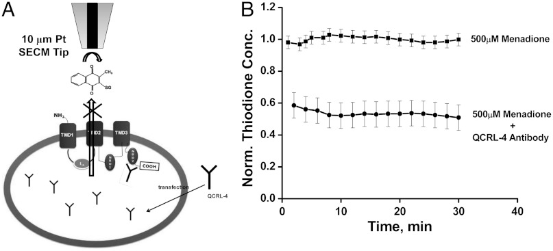
Fig. 4.
(A) Schematic diagram (not to scale) of transfection of QCRL-4 monoclonal antibody and subsequent binding to NBD2 domain of MRP1 pump. (B) Normalized thiodione concentration in presence of 500 µM menadione (upper line) without and (lower line) with antibody QCRL-4. Thiodione concentration is normalized against the average thiodione concentration produced in each individual experiment without any blocker antibody. Tip was positioned at 80 µm distance from petri dish to record the thiodione efflux.

