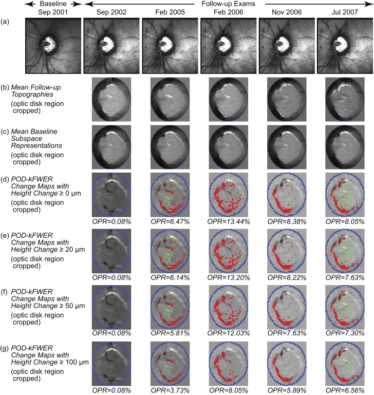
Figure 2.
POD k-FWER change significance maps of an example progressing eye. Change maps indicate locations with likely glaucomatous changes (red superpixels) and treatment effects or improvement (green superpixels). (d–g) Optic disk region is cropped for clarity. (d) The POD k-FWER detected significant glaucomatous changes (OPR >5%) in the second follow-up exam in February 2005. Application of the minimum retinal height change criterion of ≥20 μm (e), ≥50 μm (f), and ≥100 μm (g) resulted in a slight reduction in the observed positive rates. It can be noted that there was a slight reduction in OPR from 2006 to 2007, which is not reflected in the TCA maps (RC % area) due to the confirmation requirement in (Fig. 3b–e).

