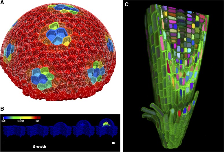
Figure 1.
Arabidopsis Serves as a Unique Plant Model for Predictive Modeling Approaches That Will Inform Attempts to Establish Similar Platforms for Crop and Ecological Research.
(A) Virtual Shoot Apical Meristem (SAM). Example of a model of the Arabidopsis SAM combining biochemical and mechanical interactions. Auxin transport model is simulated on the three-dimensional (3D) cellular template of the L1 layer of meristem. Resulting peaks of auxin are visualized as a color map from red (low) to blue (high). High auxin concentration is assumed to loosen the walls of the cells, which is simulated in a Finite Element model as lowering the elastic modulus of those walls. This leads to the pattern of the maximal principal stress direction (pictured as thin white lines) circumventing the auxin maxima, which coincide with microtubule organization during primordia formation. (Image courtesy of Henrik Jönsson, Lund University, Sweden and Elliot Meyerowitz, The Sainsbury Laboratory University of Cambridge, United Kingdom.)
(B) SAM signaling model displaying possible WUSCHEL activation by tissue geometry modifications. From left to right, a bulge grows on top of a flat tissue section. The geometrical changes are sufficient to activate the expression of WUSCHEL important for a functional shoot meristem. (Image courtesy of Henrik Jönsson, Lund University, Sweden and Elliot Meyerowitz, The Sainsbury Laboratory University of Cambridge, United Kingdom.)
(C) Virtual root. 3D visualization of an Arabidopsis root apex produced using automated image analysis and visualization techniques. Automated image analysis techniques are used to extract the 3D geometry from confocal microscope images of an Arabidopsis root apex. The extracted geometry can then be used in modeling applications to provide a foundation for realistic 3D models. (Image courtesy of the Centre for Plant Integrative Biology, University of Nottingham, United Kingdom.)

