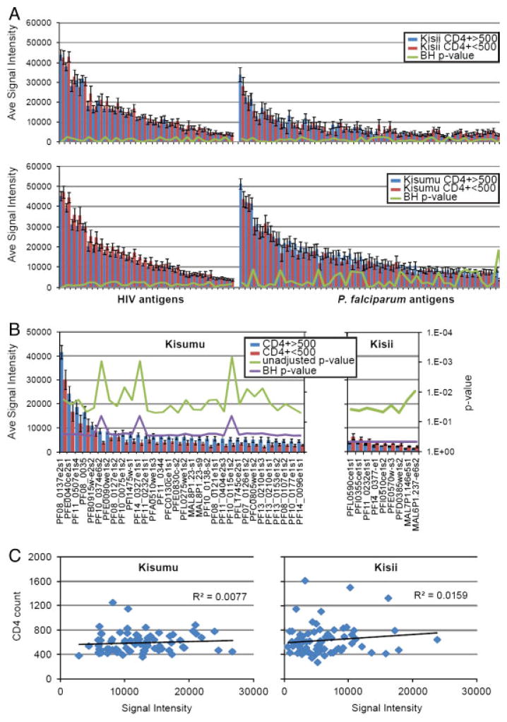
Figure 4.
Differential seroreactivity of P. falciparum in patients with low and high CD4 count. Mean seroreactivity of “low” and “high” CD4 counts are plotted in red and blue, respectively. Corresponding unadjusted p-values for each antigen are shown on the secondary axis. “e” and “s” at the end of the genes refers to exon number and segment number. (A) Comparison of the mean seroreactivity of HIV and P. falciparum antigens between low and high CD4 count individuals within Kisumu and Kisii. (B) Identification of differentially reactive antigens is displayed and the corresponding locus tags are labeled on the x-axis. (C) A scatter plot comparison of the CD4 count (y-axis) and the mean seroreactivity to the 161 seroreactive antigens (x-axis). A linear regression trend line and R2 is displayed for Kisii and Kisumu.

