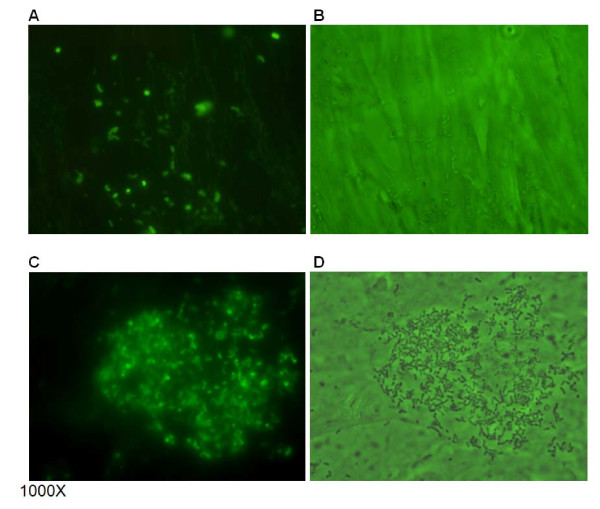
Figure 6.
Photomicrographs of bacterial adherence to bovine turbinate (BT) cells. ChoP+ and ChoP– isolates ofH. somnistrain 738 stained with a fluorescent dye adhered to BT cells. The adherence was randomly distributed per microscopic field (A and B). However, a large number of H. somni 738- were often associated with one particular cell per field (C and D) (cells with more bacteria were more commonly seen). Panels: A, fluorescence microscopy of H. somni 738+; B, mixed light and fluorescence microscopy of H. somni 738+; C, fluorescence microscopy of H. somni 738-; D, mixed light and fluorescence microscopy of H. somni 738-.

