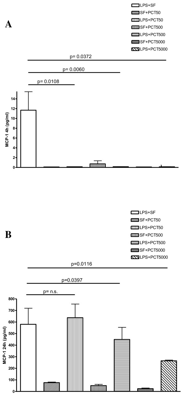
Figure 3.
In vitro effect of different concentrations of PCT on S. typhimurium LPS-induced release of MCP-1 evaluated by cytokine biochip array. Human PBMC were cultured for 4 h (panel A), and 24 h (panel B) with the following mixtures which had been pre-incubated at 37°C for 30 min : Sterile saline fluid (SF) plus 50 ng/ml PCT (SF + PCT 50); SF plus 500 ng/ml PCT (SF + PCT 500); SF plus 5000 ng/ml PCT (SF + PCT 5000); LPS of S. typhimurium SL1102 (100 ng/ml) plus SF (LPS + SF); LPS (100 ng/ml) plus 50 ng/ml PCT (LPS + PCT 50); LPS (100 ng/ml) plus 500 ng/ml PCT (LPS + PCT 500); LPS (100 ng/ml) plus 5000 ng/ml PCT (LPS + PCT 5000). Results are presented as means ± SEM of at least four experiments each carried out in duplicate. Statistical significance between groups was assessed by Student’s t test. A p < 0.05 was considered significant, whereas not significant (n.s.) difference was associated with a p ≥ 0.05. Statistics were performed in comparison with LPS-stimulated PCT-untreated cells (LPS + SF), and the exact significance index is indicated on the top of the horizontal line encompassing the two statistically compared bars

