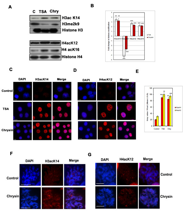
Figure 5.
Effect of chrysin on histone acetylation and methylation.(A) Histone protein expression pattern with respect to acetylation and methylation in the chrysin (40 μM) treated cells The amount of histone H3acK14, H4acK12, H4acK16 and histone H3 dimethyl lysine 9 are measured by Western blot analysis from A375 cells treated with DMSO, chrysin (40 μM) and TSA (4 μM) for 24 h. The blots are re-probed with total histone H3 and histone H4 proteins for gel loading control. (B) Bar diagrams representing the relative ratio (mean ratios ± S.D) between histone modifications upon chrysin and TSA treatment and were calculated from three independent experiments. *** represents P < 0.001; ** represents p < 0.01. (C&D) Human melanoma A 375 cells were treated with chrysin at 40 μM concentration and TSA at 4 μM concentration and the nuclei were subjected to immunostaining using H3acK14, H4acK12 antibodies. (E) Quantitative estimation of histone modifications (H3acK14 and H4acK12) in interphase nuclei in TSA and chrysin treated cells. Histogram represents quantitative measurement of histone H3acK14 and H4acK12 status as fluorescence intensity. (F&G) Metaphase spreads isolated from pre-incubated A375 cells with chrysin 40 μM and control untreated cells hybridized with histone H3acK14 and H4acK12 antibodies. Corresponding DAPI stained images of the same metaphase spreads are shown. Scale-10 μm.

