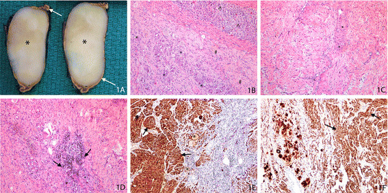
Figure 1.
Adrenal ganglioneuroma. (A) Gross photograph of the cross section of the adrenal lesion shows a stretched rim of the residual, yellow colored adrenal (arrow) surrounding a well demarcated homogenous tan-colored lesion (*). (B) Photomicrograph of hematoxylin and eosin (H&E)-stained slide at low magnification shows expansion of the adrenal medulla by the lesional cells composed of mature ganglion cells (*) admixed with Schwannian-like spindle cells (#). The residual, uninvolved normal adrenal cortex is indicated (^). (C) Photomicrograph of H&E-stained slide at high magnification shows the Schwannian-like spindle cells with wavy benign nuclei and long stretched eosinophilic cytoplasm; * highlights a focus of vague nuclear pallisading. (D) Photomicrograph of H&E-stained slide at low magnification shows collections of benign lymphocytes (arrow) in a neurotropic distribution along the mature ganglion cells (*). (E) Photomicrograph of immunohistochemically-stained slide of S100 at low magnification shows the Schwannian-spindle cells (arrow) and the ganglion cells (*) to be strongly positive. (F) Photomicrograph of immunohistochemically-stained slide with neurofilament at low magnification shows the Schwannian-spindle cells (arrow) and the ganglion cells (*) to be strongly positive.

