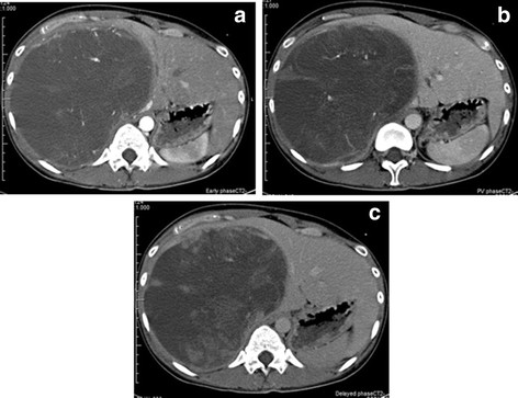
Figure 1.
(a, b, and c) Abdominal computed tomography scan showing a well-defined, low-density large mass (21 × 19 × 14 cm) with heterogeneity in the right segments of the liver. This tumor had vessels, septa, no enhanced-effect, and no solid compartment (a, early phase; b, portal vein phase; and c, delayed phase).

