Figure 2.

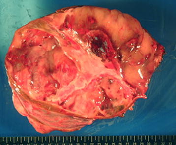
The cut surface showing a grayish-red soft mass with cystic degeneration, together with areas of hemorrhage and necrosis.

The cut surface showing a grayish-red soft mass with cystic degeneration, together with areas of hemorrhage and necrosis.