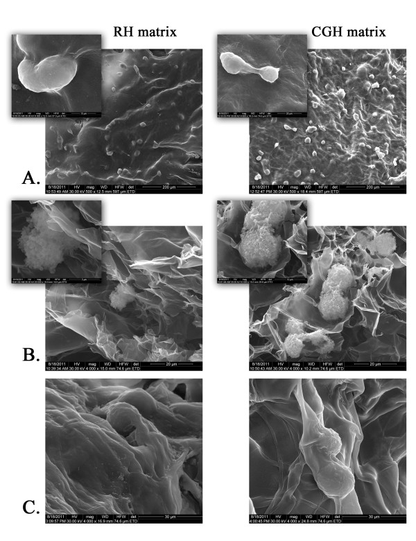
Figure 3.
Scanning Electron Microscopy (SEM) micrographs of both 3-D porous cell-laden RH and CGH matrices. A) Surface appearance of RH and CGH matrices after 2 days of culture; B) Cross-sections of the cell-laden 3-D structures at 2 days post-seeding; C) Cross-sections of cell-laden hydrogels at 7 days post-seeding.

