
Figure 4. N-terminal helix of Nef positions the Nef core domain near the membrane for efficient interactions with MHC-I and μ1.
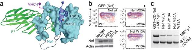
(a) Nef N-terminal helix (blue) is anchored to the core (cyan) via two hydrophobic residues, Trp13 and Met20. (b,c) Mutagenesis verification in vivo (FACS) and in vitro (GST pull-down assay), respectively, of the importance of the two residues. FACS experiments were carried out using human T lymphocytes of cell line SupT1 transfected to express Nef and GFP (as a transfection marker), followed by staining for surface MHC-I. Relative fluorescence intensity of MHC-I versus GFP. Nef, red; negative control (GFP only), blue. Lower left in b, western blots of Nef proteins.