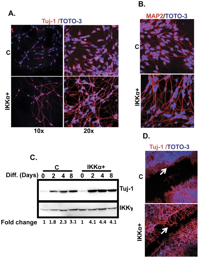
Figure 2. Elevated IKKα promotes the differentiation of MESC2.10 cells.
(A, B) IKKα promotes neurite outgrowth in differentiating NPCs. Control (C) and IKKα+NPCs were differentiated on coverslips for 4 days, fixed and stained with neuronal differentiation markers Tuj-1 (A) and MAP-2 (B). Representative micrographs obtained with a confocal microscope are shown. The DNA stain TOTO-3 was used to identify nuclei. (C) Tuj-1 levels are elevated in differentiating IKKα+ NPCs. Representative western blot results are shown for cytoplasmic lysates staining for Tuj-1 levels at different time points during the differentiation of control and IKKα+ NPCs [diff.(days)]. IKKγ was used as a loading control. Fold-change was obtained by dividing the intensity of Tuj-1 to the corresponding IKKγ, obtained by a Fluorchem 8900 (Alpha Innotech, San Leandro, CA). (D) The scratch assay shows extensive neurite outgrowth in differentiating IKKα+ NPCs. Cultures on the 2nd day of differentiation were wounded by a micropipette tip and further incubated for additional two days. Cells were fixed and stained as above. Arrows point to the areas of neurite extension.

