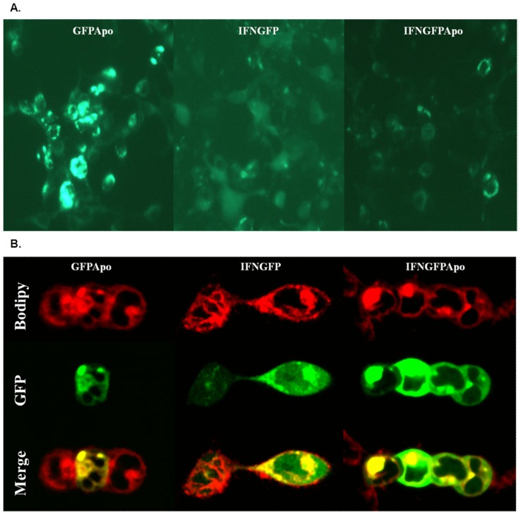
Figure 1. Evaluation of GFP activity by microscopy analysis.
2×10⁵cells per well were seeded and transfected with pGFPApo, pIFNGFP and IFNGFPApo complexed in PEI 25000(kDa). 24 hours after transfection of GFPApo (left), IFNGFP (center) and IFNGFPApo (right) A. fluorescence was evaluated by fluorescence microscopy or B. cells were dyed with BODIPY TR and analyzed by confocal microscopy.

