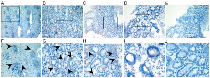
Figure 1. Immunohistochemistry with FcεRIα specific antibody (mAb 15-1) on snap-frozen intestinal specimens from (A) the esophagus, (B) the stomach, (C) the duodenum, and (D) the colon.
FcεRIα-positive cells (red) are frequently found in the esophagus, the stomach, and the duodenum (black arrows). (E) shows isotype control with mouse IgG1. Goblet cells in the duodenum and the colon revealed non-specific binding of antibodies. Original magnification x20. Bottom row (F-J) shows details from A-E. Representative specimens from n = 10.

