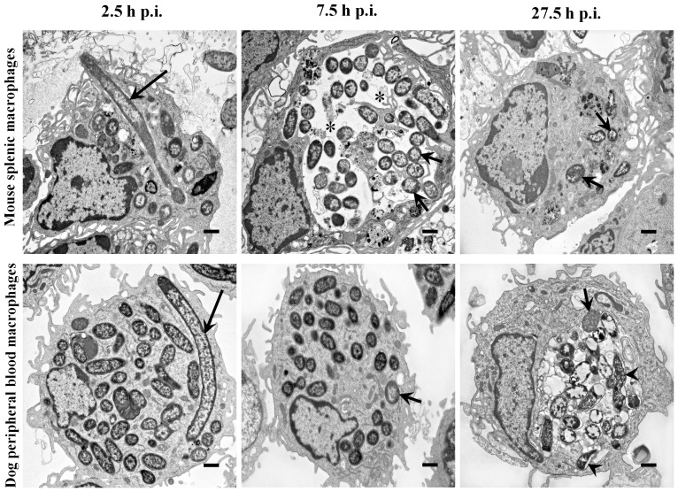
Figure 2. Ultrastructural features of intracellular Y. pestis and infected mouse and dog primary macrophages.
Primary macrophages from mouse spleen (top row) and dog peripheral blood (bottom row) infected with Y. pestis strain KIM6+ were glutaraldehyde fixed at 2.5, 7.5 and 27.5 h p.i. and examined using TEM. Long arrows, short arrows and arrow heads represent filamentous, coccobacillary and degraded coccobacillary Y. pestis, respectively. Asterisks indicate spacious vacuolar extension of phagolysosomes. The images are at a 6,000× magnification, and the cross bars represent 1 µm.

