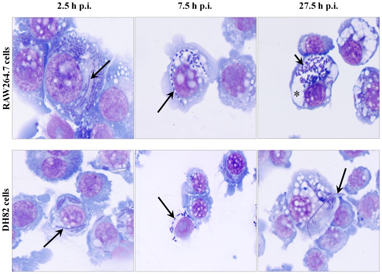
Figure 4. Morphological features of intracellular Y. pestis and of infected mouse and dog tissue culture cells.
Y. pestis strain KIM6+ infected RAW264.7 (top row) and DH82 (bottom row) cells were sampled from 2.5 to 27.5 h of p.i. and observed under light microscope by staining with Wright Giemsa stain. Filamentous and coccobacillary forms of intracellular Y. pestis and spacious phagolysosomal extensions were determined at a 1,000× magnification. Long and short arrows indicate filamentous and coccobacillary form of intracellular Y. pestis, respectively. Asterisk indicates spacious vacuolar extension of phagolysosome.

