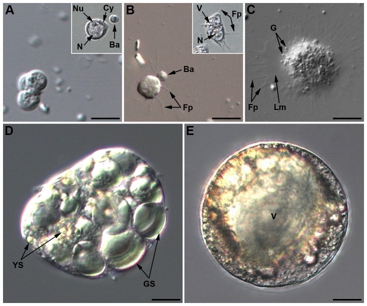
Figure 1. Light microscopy pictures of Acyrthosiphon pisum hemocytes (LL01 clone).
(A) Three prohemocytes in cluster. Inset: phase contrast showing the large central nucleus and the nucleolus (Nu). Ba: B. aphidicola. (B) Plasmatocyte beginning to adhere, with filopodia (Fp) extension. Inset: phase contrast showing large cytoplasmic vacuolar formation. (C) Adherent granulocyte containing cytoplasmic granules (G) and filopodia (Fp) extending from a lamellipodium (Lm). (D) Spherulocyte with its large colored globular inclusions, small yellow spherules (YS) and large green spherules (GS). (E) Wax cell showing a large central vacuole (V) and colored globular inclusions that differ from those of spherulocytes. Same magnification for all micrographs; scale bar: 10 µm.

