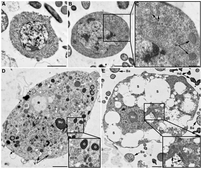
Figure 2. TEM characterization of hemocytes.
Cells from the LL01 clone (A–D) and the YR2-Hd line (E). (A) Prohemocyte characterized by a round nucleus (N), a high N/C ratio, and homogenous cytoplasm devoid of apparent organelles. (B) Plasmatocyte with homogeneous cytoplasm, a lobulated nucleus, a high N/C ratio and some cytoplasmic organelles clearly visible in box (C): Rough Endoplasmic Reticulum (RER), Mitochondria (M). (D) Granulocyte with a lobulated nucleus, a low N/C ratio, and granules (G). The cytoplasm contains numerous organelles (see box). A phagosome is observed that contains a large foreign particle (asterisk). (E) YR2-Hd spherulocyte with a round nucleus and a low N/C ratio. The large volume of cytoplasm is filled with spherules (S) of different sizes, and numerous mitochondria are found in a small region (enlarged box). Ba: B. aphidicola; Hd: H. defensa. Scale bar: 2 µm (A, B, D, E) and 0.5 µm (C).

