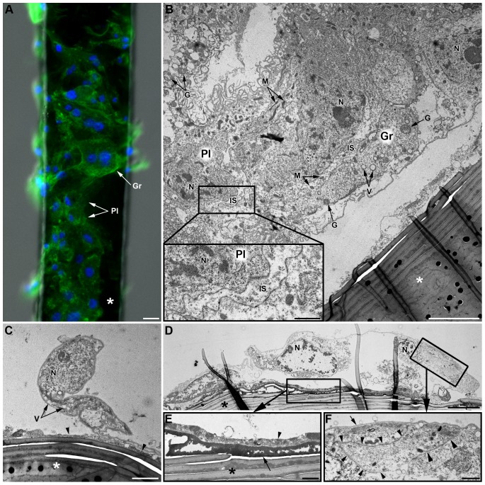
Figure 6. In vivo hemocyte adhesion assay (LL01 clone).
(A) Observations of a brush horsehair (asterisk) seven days after insertion in the hemocoele of an LL01 aphid: both plasmatocytes (Pl) and granulocytes (Gr) are observed (F-actin (green) and nuclei (blue) staining). Scale bar: 10 µm. (B–F) TEM micrographs of the “encapsulation” process. (B) Part of a multilayered hemocytic capsule containing both plasmatocytes (Pl) and granulocytes (Gr). The capsule is partly detached from the brush hair (asterisk). Intercellular spaces (IS) between hemocytes are relatively large (see box magnified bottom left) and they often contain some cytoplasmic organelles (mitochondria, granules…). Scale bar: 5 µm; box: 1 µm. (C) Plasmatocyte partly adhering to the brush hair (asterisk). An electron-dense matrix of granular aspect is observed at the interface between the hair and the hemocyte (arrowhead). Scale bar: 2 µm. (D) Granulocyte largely spread onto the hair. Two characteristic layers are observed at the interface between the horsehair and the hemocyte (box magnified in (E)). Scale bar: 5 µm. (E) The internal layer is highly electron-dense, compact and homogenous (arrow). The external layer is heterogeneous and composed of cell debris and coagulated hemolymph (arrowhead). Scale bar: 1 µm. This granulocyte is in tight contact with another “capsule”-forming hemocyte (box magnified in (F); arrowheads). An electron-dense thin layer covers the surface of the cell (arrow). Scale bar: 1 µm. N: Nucleus; M: Mitochondria; G: Granule; V: Vacuole.

