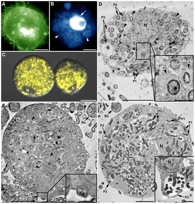
Figure 8. In vivo phagocytosis of symbionts by hemocytes from SS-containing YR2 lines.
(A–C) Fluorescent micrographs of YR2(Ri) hemocytes. (A) F-actin (green) and (B) DNA staining (blue), of an adherent granulocyte. The cytoplasm is filled with R. insecticola secondary symbionts as shown by bacterial DNA staining (arrowheads in B; arrow: cell nucleus), and (C) specific FISH detection (yellow, false color; DIC and confocal micrographs overlay). Nucleus (N) location is detectable by the absence of yellow coloration. (D–F) TEM micrographs showing phagocytosis of secondary symbionts by hemocytes of three YR2 lines. (D) YR2(Ri) granulocyte containing numerous R. insecticola inside the cytoplasm. Different symbiont-containing phagosomal structures are observed (P1: zipper-like, P2: trigger-like, P3: macropinocytosis), as well as phagolysosomes containing symbionts and membranous material (arrowheads and box). (E) YR2-Ss line granulocyte with numerous S. symbiotica inside cytoplasmic membranous phagolysosomes (arrowheads). Insert box: detail of S. symbiotica engulfment by trigger-like phagocytosis. (F) YR2-Hd granulocyte containing H. defensa symbionts in the three types of phagosomes. Inset: plasmatocyte with a large phagosome structure (macropinocytosis-like) enclosing several symbionts and extracellular fluid. Scale bar: 5 µm (A–B), 10 µm (C), 5 µm (D), 2 µm (F) and 1 µm (magnifications boxes in (D) and (E)).

