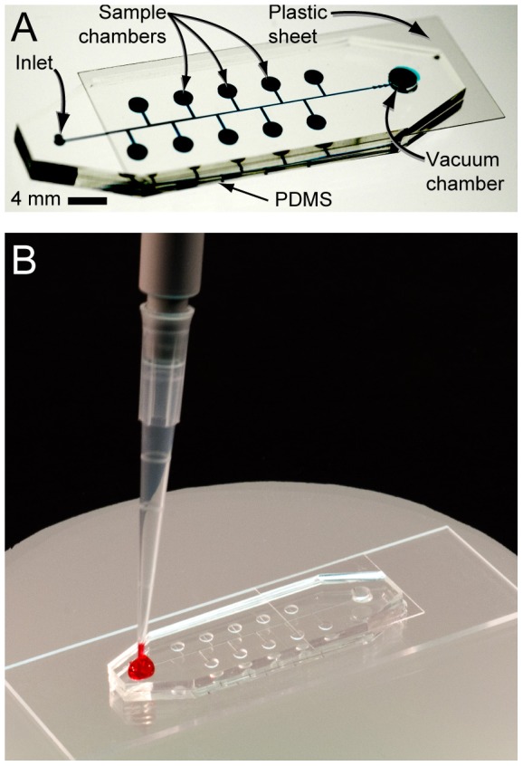
Figure 2. Images of the BacChip.

The device consists of 10 microchambers; 9 of the microchambers are preloaded with a different saccharide and one is left empty as a control. We punched holes at the ends of the center PDMS channel to form an inlet port and a vacuum chamber. The PDMS layer was pressed into contact with a glass slide to form the microfluidic system. A plastic sheet was placed over the vacuum chamber to seal it. After removing the device from vacuum it is ready for sample loading by the user. (A) An image of an assembled device in which the channels and chambers are filled with a blue dye to make them visible. (B) After removal from vacuum, the user adds 20 µL of sample to the inlet port and the device fills and isolates cells in the microchambers autonomously.
