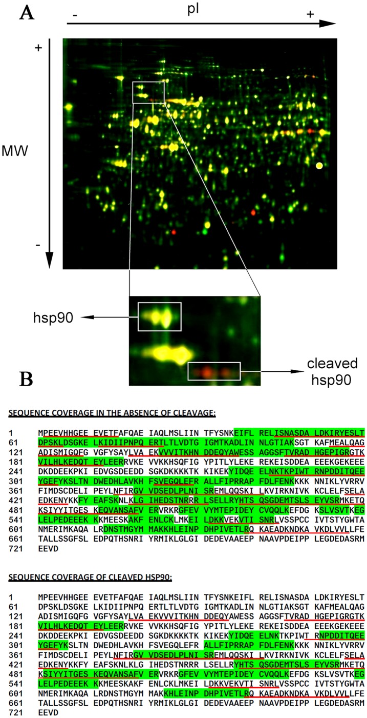
Figure 3. 2D-DIGE analysis of protein extracts from K562 cells exposed to oxidative stress.
(A) K562 cells incubated for 2 h in the absence (green) or the presence (red) of A/M (2 mM/10 µM). Spots corresponding to Hsp90 and its cleaved fragments were excised and analyzed by mass spectrometry. (B) Peptides found after mass spectrometry analysis of the excised 2D-DIGE spots of cleaved and non-cleaved Hsp90 protein by either tryptic (highlighted in green) or chymotryptic (underlined in red) peptides. Alignment was performed against the Hsp90β protein sequence.

