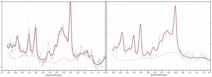
Figure 2. Raw (light gray), fit (heavy solid red), and baseline (solid gray) LCModel (Provencher 2001) output spectra for proton magnetic resonance spectroscopy voxels placed in midline pregenual anterior cingulate (pACC) in study subject with ASD (left) and control subject (right).
Note larger Glx peak (2.0–2.5 ppm) in subject with ASD. Seven of 8 subjects with ASD exhibited Glx levels above the control mean (Fig. 3). Experiment 1.

