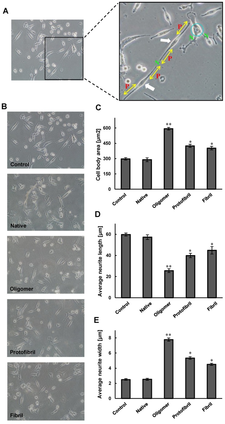
Figure 4. The effects of oligomer, proto-fibril and fibril forms of insulin on neurite outgrowth in differentiated PC12 cells.
A) The criteria of PC12 differentiation are shown on three neurons (left image) of a sample image. The “P” on right image indicates the primary neuritis of a neuron. The yellow arrow shows the length of a neurite, extent elongated, and membrane-enclosed protrusions of cytoplasm. The blue circle on right image shows the cell body. Neurite width is not equal in all parts of neurons, thus the average neurite width must be calculated by dividing cell body area to average neurite length. The white arrows show to bipolar cells. The letter “N” indicates the nodes, the sites at which individual neurites branched or separate neurites contacted each other. The criteria were quantified 12 h after treatment; B) NGF-differentiated PC12 cells were pretreated with three amyloid intermediate forms of insulin. C) Cell body area; D) average neurite length; and E) average neurite width. *Significantly different from control cells. Statistical significances were achieved when p<0.05 (*p<0.05 and **p<0.01).

