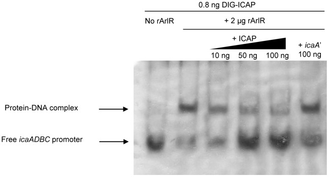
Figure 7. Binding of ArlR to the icaADBC promoter.
Lanes were loaded as follows: lane 1, 0.8 ng Dig-ICAP alone; lane 2, 0.8 ng Dig-ICAP and 2 µg rArlR; lanes 3–5, Dig-ICAP, rArlR and increasing amounts of unlabeled ICAP (12.5, 62.5,125 fold increase in Dig-ICAP, respectively); and lane 6, Dig-ICAP, rArlR and 100 ng of a 405 bp icaA fragment. The DIG-labeled DNA fragments were transferred to positively charged nylon membranes and visualized by an enzyme immunoassay using anti-Digoxigenin-AP, Fab-fragments and the chemiluminescent substrate CSPD. Chemiluminescent signals were recorded on X-ray film. DIG-ICAP, digoxin-labeled icaADBC promoter region; ICAP, unlabeled icaADBC promoter region; icaA’, icaA gene fragment.

