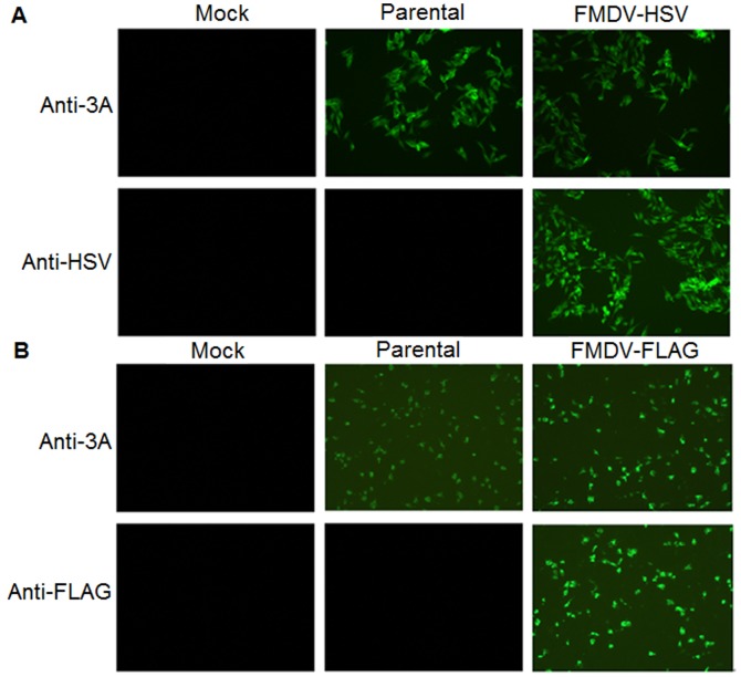
Figure 2. Immunofluorescence analysis of the expression of epitope tags in recombinant viruses.
Confluent BHK-21 cells were mock-infected or infected with r-HN or the transfected supernatants at a MOI of 1, incubated for 6 h, fixed and probed with anti-3A and anti-HSV (A) or anti-FLAG mAb (B), followed by incubation with FITC-conjugated secondary antibody. The cells were visualized under an Olympus BX40 fluorescence microscope. Magnification, ×10.

