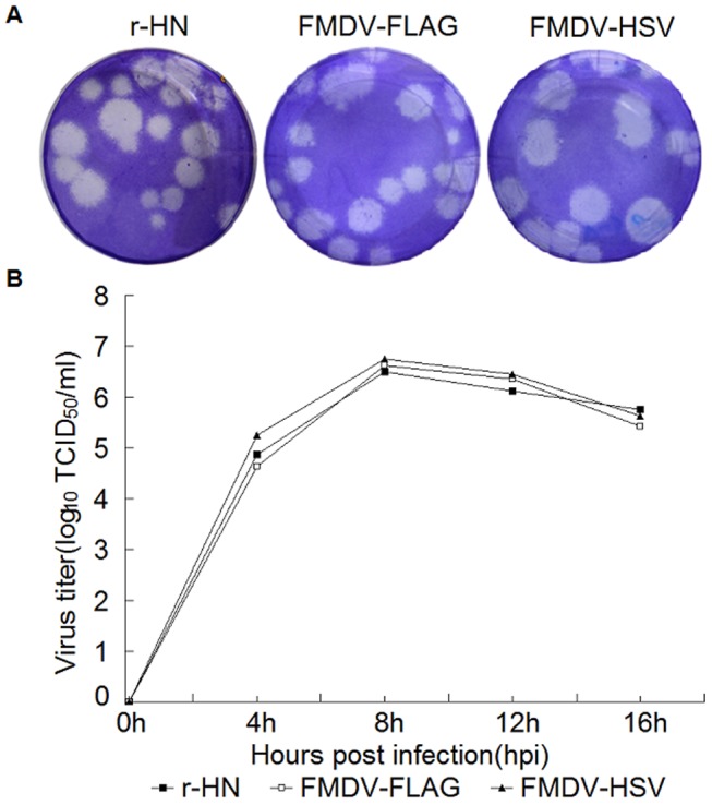
Figure 5. Plaque morphology and growth curves of the parental virus and the 3A-tagged viruses in BHK-21 cells.

(A) Plaque morphology of the parental virus and the 3A-tagged viruses in BHK-21 cells. Viruses were plaque-assayed under tragacanth gum on BHK-21 cells and cells were stained at 48 h hpi. Plaques from appropriate dilutions are shown. (B) Growth curves of the parental virus and the 3A tagged viruses in BHK-21 cells. BHK-21 cells were infected with the parental virus or the 3A-tagged viruses at a MOI of 1. At 0, 4, 8, 12 and 16 hpi, cells and supernatants were harvested and virus titers were determined by TCID50/ml on BHK-21 cells. The values of the viral titer represent the average obtained from triplicate experiments.
