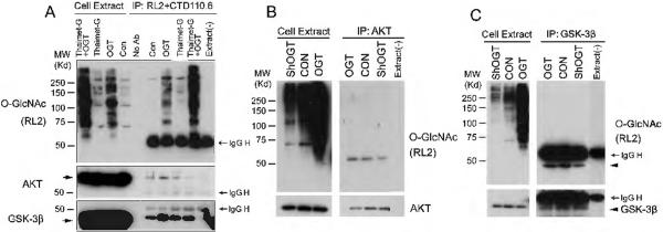
Fig. 1.
Detection of O-GlcNAcylation of AKT and GSK-3β. (A) HEK-293FT cells were transfected with OGT for 36 hr and then treated with 25 μM thiamet-G for 12 hr. The OGlcNAcylated proteins were immunoprecipitated from the cell lysate using a mixture of two antibodies, RL2 and CTD110.6, and detected by Western blots developed with RL2. (B,C) AKT (B) and GSK-3β (C) were immunoprecipitated from the lysates of HEK-293FT cells after transfection with OGT or shOGT for 48 hr, and O-GlcNAcylation level was detected by Western blots developed with RL2. As a control, a buffer instead of cell extracts was added(right lane). The AKT (B) and GSK-3β (C) blots were included to confirm the success of immunoprecipitation.

