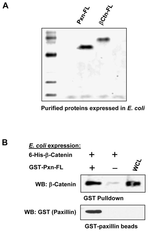
Figure 3. Interaction between paxillin and β-catenin expressed in E. coli.
A: Full length paxillin and β-catenin were expressed in E. coli (strain BL21-AI). Fusion proteins were purified using GSH-sepharose and Ni-column, respectively, and analyzed by SDS-PAGE followed by Coomassie staining. B: 6-His-tagged β-catenin and full length GST-tagged paxillin expressed in the bacterial system were tested in a GST-pulldown assay. GST conjugated to the beads alone served as negative control. After washing steps, beads were treated with SDS sample buffer, and bound paxillin and β-catenin were determined by western blot with corresponding antibody.

