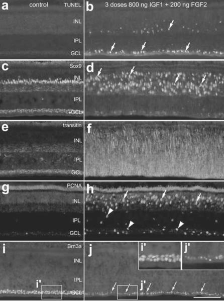
Figure 3.
Treatment with 3 consecutive daily injections of 800ng IGF1 and 200ng FGF2 causes cell death, glial migration and proliferation within the retina. Retinas were harvested 1 day after the last of 3 consecutive daily injections of factors (paradigm 1). Vertical sections of central regions of the retina were labeled for fragmented DNA using the TUNEL method (a and b) or with antibodies to Sox9 (c and d), transitin (e and f), PCNA (g and h) or Brn3a (I and j). Arrows indicate dying cells (b), reactive Müller glia (d), proliferating Müller glia (h) or abnormal ganglion cell nuclei (j). Arrow-heads indicate putative NIRG cells that are PCNA-positive (h). The insets (i' and j') in panel j are 2-fold enlargements of the boxed-out regions in i and j. The calibration bar (50 μm) in panel j applies to all panels. Abbreviations: INL – inner nuclear layer, IPL – inner plexiform layer, GCL – ganglion cell layer, PCNA – proliferating cell nuclear antigen.

