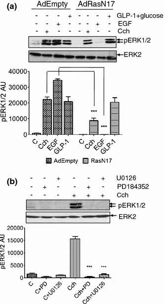
Fig. 2.

Role of Ras and MEK in carbachol-stimulated ERK1/2 activation. a MIN6 cells were infected with a control adenovirus (AdEmpty.eGFP) or with an adenovirus engineered for expression of RasN17 (AdRasN17). After 48 h, cells were pre-incubated for 1 h with KRB-minus-glucose before treatment with carbachol (1 mM, 2 min), EGF (20 ng/ml, 5 min) or GLP-1 (10 nM) plus 16.7 mM glucose for 10 min. b MIN6 cells were pre-incubated for 1 h in KRB and where indicated PD098059 (PD, 1 μM) or U0126 (20 μM) was added for the last 30 min before addition of carbachol (1 mM, 2 min). Proteins were separated by SDS–PAGE and detected by Western blotting using anti-phospho-ERK1/2 and anti-ERK2 antibodies. Below each representative blot is a graph showing densitometric quantification of ERK1/2 phosphorylation. Data are presented as means SEM (n = 3); ***P < 0.001 by Dunnett’s range test following one-way ANOVA compared to the carbachol response
