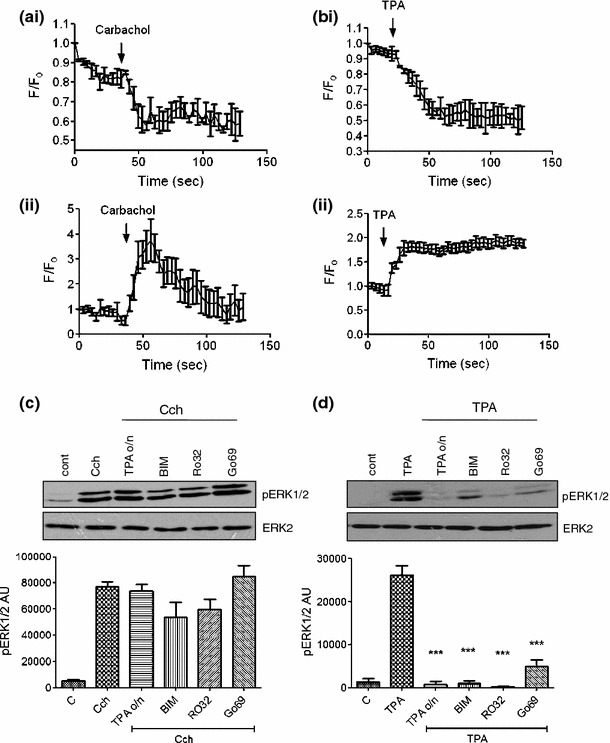
Fig. 3.

PKC requirement for carbachol-mediated ERK1/2 activation. MIN6 cells, transfected to express eGFP-tagged PKCε, were pre-incubated for 1 h in KRB-minus-glucose prior to treatment with a carbachol (1 mM) or b TPA (1 μM). Fluorescence intensity in the (i) cytoplasm or (ii) at the membrane was monitored by fluorescence confocal microscopy. Changes in fluorescence intensity were expressed relative to initial fluorescence (F/F 0). c, d MIN6 cells pre-treated in the absence or presence of TPA (1 μM) for 16 h (TPA o/n) were pre-incubated for 1 h in KRB-minus-glucose prior to treatment with c carbachol (1 mM, 2 min) or d TPA (1 μM, 60 min) in the absence or presence of bisindolylmaleimide I (BIM, 1 μM), Ro 32-0432 (Ro32, 1 μM), or Gö6976 (Go69, 1 μM). Proteins were separated by SDS–PAGE and detected by Western blotting using anti-phospho-ERK1/2 and anti-ERK2 antibodies. Representative blots are shown above with mean data densitometry below in panels c and d. Data are shown as means + SEM (n = 3); ***P < 0.001 by Dunnett’s range test following one-way ANOVA compared to carbachol (panel c) or TPA (panel d)
