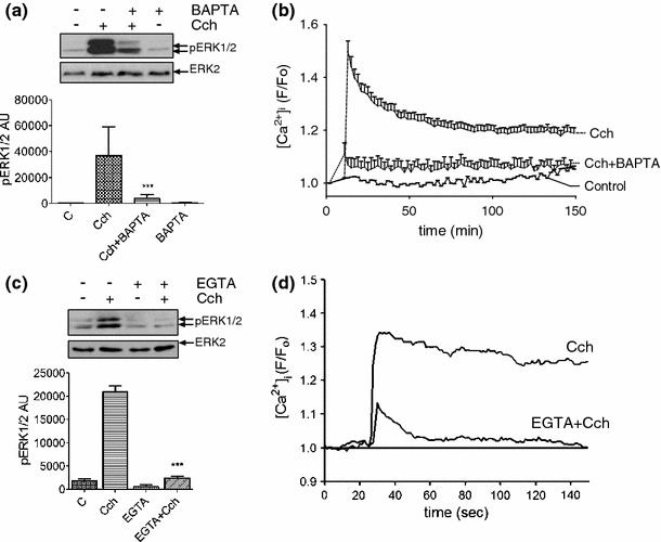
Fig. 4.

Ca2+ influx across the plasma membrane is required for carbachol-stimulated ERK1/2 activation. a MIN6 cells were pre-incubated for 30 min in KRB-minus-glucose and, where indicated, loaded with BAPTA-AM (100 μM, BAPTA) for 30 min. Cells were then treated with carbachol (1 mM, 2 min). Proteins were separated by SDS–PAGE and detected by Western blotting using anti-phospho-ERK1/2 and anti-ERK2 antibodies. A representative blot is shown above with mean data densitometry below. Data are shown as means SEM (n = 3); ***P < 0.001 by Dunnett’s range test following one-way ANOVA compared to carbachol. b Cells were loaded with Fluo-4-AM (2 μM) at the same time as BAPTA-AM and [Ca2+]i measured using single-cell confocal Ca2+ imaging following carbachol (1 mM) addition. Data represent means SEM for the increase in [Ca2+]i levels (n > 30). c MIN6 cells were pre-incubated for 30 min in KRB-minus-glucose and, where indicated, incubated in EGTA-buffered KRB for the last 10 min. The Ca2+ concentration in the KRB was reduced to ≤ 100 nM by EGTA-buffering and confirmed using Fura-2 free acid and standard fluorimetry. Carbachol (1 mM) was added to the KRB for 2 min. Proteins were separated by SDS–PAGE and detected by Western blotting using anti-phospho-ERK1/2 and anti-ERK2 antibodies. A representative blot is shown above with densitometry below. Data are means SEM (n > 3); ***P < 0.001 by Dunnett’s range test following one-way ANOVA compared to carbachol. d Changes in [Ca2+]i were assessed by single-cell confocal Ca2+ imaging reproducing the experimental conditions used in panel c
