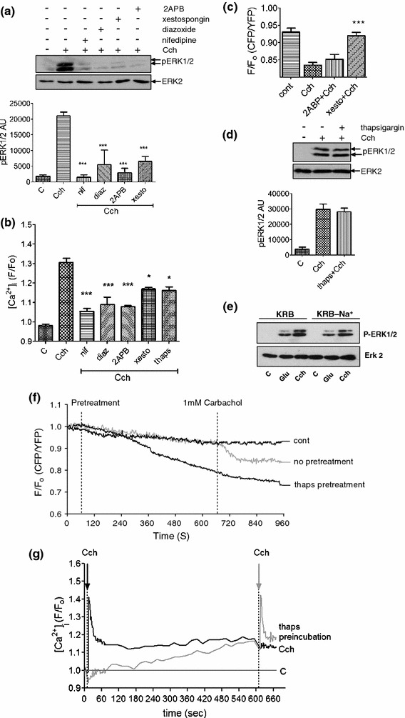
Fig. 5.

Multiple Ca2+ sources are required for carbachol-stimulated ERK1/2 activation. a MIN6 cells were pre-incubated for 1 h with KRB-minus-glucose in the absence or presence of 2APB (10 μM) for 30 min, or nifedipine (10 μM), xestospongin C (10 μM) or diazoxide (250 μM) for 10 min prior to carbachol (1 mM, 2 min) addition. Proteins were separated by SDS–PAGE and detected by Western blotting using anti-phospho-ERK1/2 and anti-ERK2 antibodies. A representative blot is shown above with mean data densitometry below. Data are shown as means SEM (n = 3); *P < 0.05; ***P < 0.001 by Dunnett’s range test following one-way ANOVA compared to carbachol addition. b [Ca2+]i was monitored in populations of MIN6 cells pre-treated as in a by NOVOstar platereader. Data are shown as means SEM (n = 3); *P < 0.05; ***P < 0.001 by Dunnett’s range test following one-way ANOVA compared to carbachol addition. c MIN6 cells transfected with the cameleon D1ER construct were pre-incubated for 1 h in KRB-minus-glucose. Cells were then incubated in the absence or presence of 2ABP (10 μM) or xestospongin C (10 μM) for 10 min before the addition of carbachol (Cch, 1 mM). Data shown represent peak FRET changes after agonist addition. d MIN6 cells were treated as in a, but in the absence or presence of thapsigargin (1 μM) pre-addition for 10 min. A representative blot is shown above with mean data densitometry below. e MIN6 cells were maintained in KRB in which Na+ was replaced by methylglucamine for 15 min before stimulation with either glucose (20 mM) or carbachol (1 mM). Data are shown as means SEM (n = 3); *P < 0.05; ***P < 0.001 by Dunnett’s range test following one-way ANOVA compared to carbachol. f MIN6 cells transfected with cameleon D1ER were pre-incubated for 1 h in KRB-minus-glucose prior to recording. All recordings show 1 min of basal KRB perfusion before either no pre-treatment or thapsigargin (1 μM) addition for 10 min followed by the addition of carbachol (1 mM) for 5 min. Data shown represent mean changes in FRET for n ≥ 30 cells. g MIN6 cells were pre-incubated in KRB-minus-glucose and then treated with carbachol (1 mM, black line) or thapsigargin (1 μM, gray line) for 10 min followed by the addition of carbachol (1 mM). Fluorescence was measured as an indicator of [Ca2+]i by NOVOstar platereader for 10 s before initial Cch/thapsigargin pre-treatments
