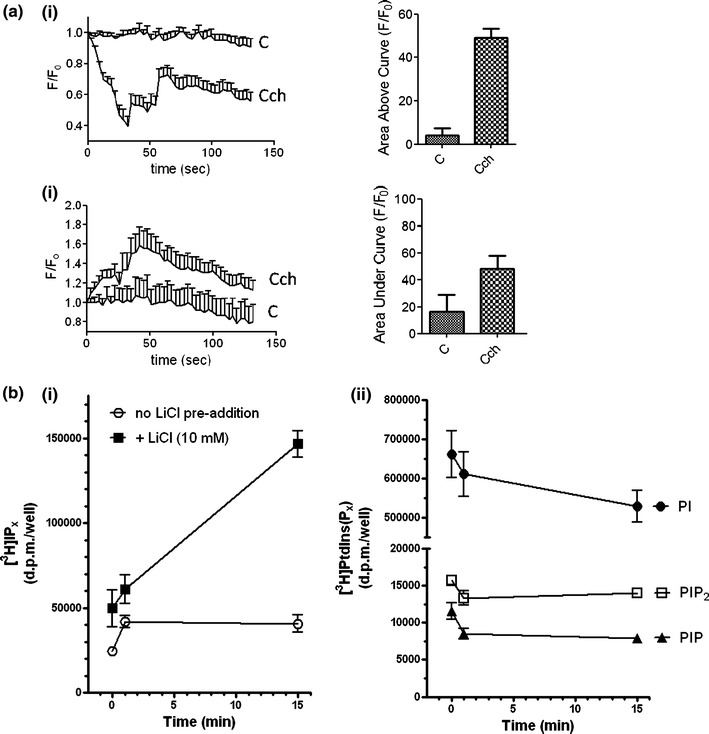
Fig. 6.

Changes in phosphoinositide turnover in MIN6 cells in response to carbachol. a MIN6 cells were transfected with a plasmid encoding eGFP-PHPLCδ1. Cells were then pre-treated for 1 h with KRB-minus-glucose and treated with carbachol (1 mM). Changes in fluorescence intensity at the plasma membrane (i) or in the cytoplasm (ii) were determined by confocal microscopy. Fluorescence intensity was referenced to the initial fluorescence (F/F 0). The area under the baseline for membrane fluorescence or over the baseline for cytoplasmic fluorescence was calculated and compared between control and carbachol-stimulated cells. Data are presented as means SEM (n > 30). b MIN6 cells were incubated with [3H]inositol for 48 h. Cells were pre-incubated for 1 h with KRB-minus-glucose and, where indicated, LiCl (10 mM) was added for the final 30 min. Cells were challenged with carbachol (1 mM) for the times indicated and [3H]IPx and [3H]PtdIns(Px) fractions recovered and analyzed as described in the “Methods” section. Time-dependent effects of carbachol stimulation on (i) [3H]IPx accumulations in the absence and presence of LiCl and (ii) [3H]PtdIns, [3H]PtdInsP and [3H]PtdInsP2. Data are shown as means + SEM for 3 independent experiments performed in duplicate
