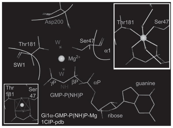
Figure 4.
Architecture of the Mg-binding pocket in GTP-Gi1α. Upper inset: green lines: coordination bonds of Mg; light blue lines: hydrogen bonds. Note that electrons of six oxygens coordinate Mg: one from the β phosphate of GTP, one from the γ phosphate of GTP, one from the hydroxyl of Ser47, one from the hydroxyl of Thr181, and two from a for Mg water molecules stabilized by hydrogen bonds to Asp200 and to the α phosphate of GTP. Lower Inset: octahedral coordination cage.

