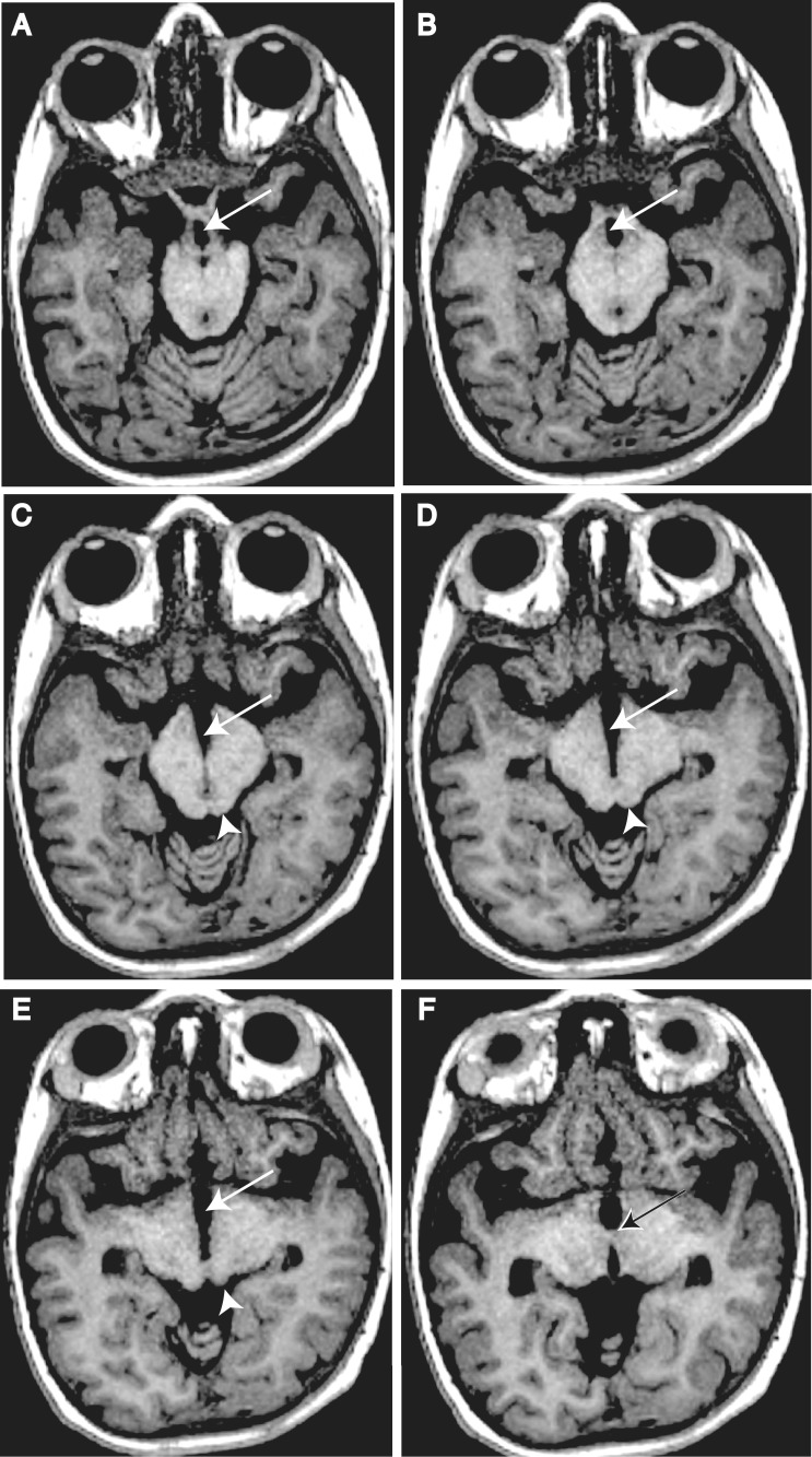
Figure 3.
Key anatomical features of DMJD. T1-weighted axial 1.2 mm thin sections from Patient DMJD-1592-2, demonstrating that the anterior midbrain cleft is continuous with the third ventricle. (A and B) Arrows indicate chiasmatic recess. (A) Optic track entering the midbrain. (B) Optic radiations coursing around midbrain. (C and D) Arrows indicate third ventricle extending posteriorly, producing the ‘butterfly’ sign. Arrowhead indicates inferior colliculi. (E) Arrow indicates third ventricle. Arrowhead indicates superior colliculi. (F) Arrow indicates massa intermedia (interthalamic adhesion), defining posterior limit of third ventricle.

