Figure 5.
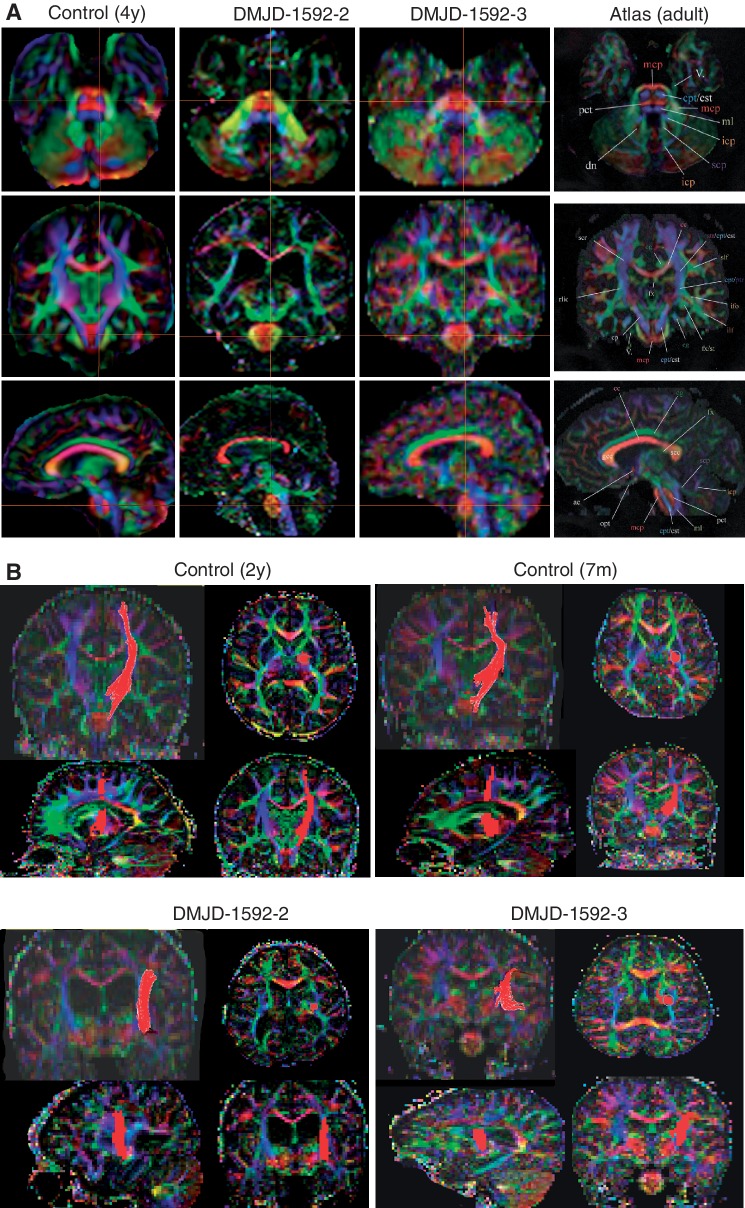
Disrupted corticospinal tract in DMJD. (A) Multiplane reconstruction with directional encoded colours from diffusion tension imaging of paediatric control, and two affected members in Family DMJD-1592 in comparison with MRI atlas of human white matter (Mori et al., 2011). Top: Axial plane in control shows corticopontine/corticospinal tracts evident in pons in blue (crosshairs), whereas neither patient has evident corticopontine/corticospinal tract. Middle: Coronal plate also shows corticopontine/corticospinal tract at crosshairs in control but absent in both patients. Bottom: Parasagittal plane also shows evident corticopontine/corticospinal tract in control, absent in patients. Crosshairs represent the relative plane of imaging for each of the images in the series for maximal registration correspondence. (B) Region of interest tractography of corticopontine/corticospinal tract fibre bundle (red) in each orientation from aged-matched control and the two affected members of Family DMJD-1592. Control shows the usual course of corticopontine/corticospinal tract, originating in the cortical motor area, lateral to the ventricle and then heading medially in the diencephalon/mesencephalon. Both patients show aberrant course of corticopontine/corticospinal tract, coursing laterally.

