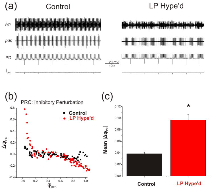
Figure 3.
The LP to PD synapse attenuated the disturbing influence of extrinsic random perturbations. a. Extracellular recording traces from the nerves lvn and pdn and intracellular voltage traces from the PD neuron indicate the activity of pyloric circuit neurons. A brief current pulses (−2 nA, 50 ms) were injected into the PD neuron every 10 seconds for 15 minutes in the presence of the LP to PD synapse (Control) or when the LP neuron was functionally removed by hyperpolarization (LP Hype’d). b. An example of PRC in response to the inhibitory perturbation (−2 nA, 50 ms). The phase reset (ΔϕPD ((P0 – P) / P0)) was plotted against the perturbation phase ϕpert (Δt / P0). The Inhibitory perturbation shortened the period at the early phases and prolonged the period at late phases. In the presence of the LP to PD synapse the PRC (ΔϕPD) lies closer to zero compared to LP Hype’d. c. Comparison of the mean PRC values between Control and LP Hype’d. When the LP neuron was functionally removed (LP Hype’d) the mean of the absolute value of ΔϕPD (|(P0 – P) / P0)|) was significantly larger compared to control in response to the perturbations (Student’s t-test, p<0.001; N=4 preparations, n=178 stimuli per preparation).

