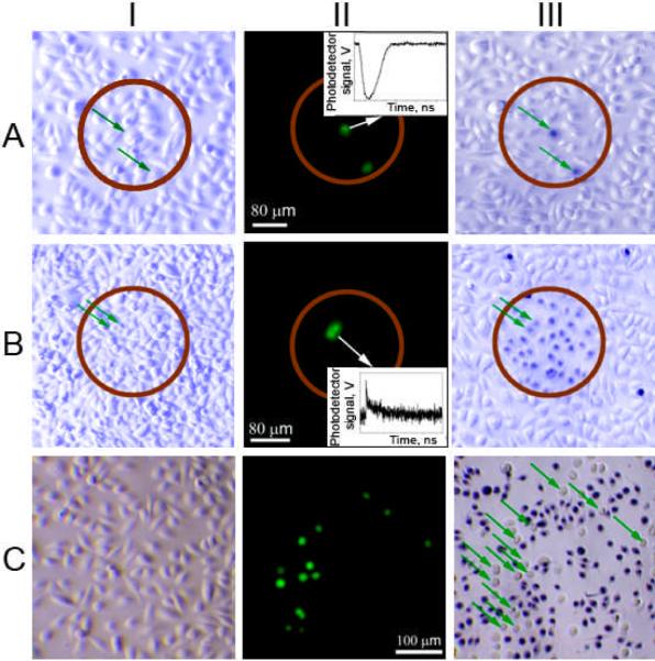
Figure 2.

Bright field (I, III) and fluorescent (II) microscopy images of a co-culture of normal (NOM9) and squamous cell carcinoma (HN31, green or shown with green arrows) cells, the images I and II were taken before treatment and the image III was taken after the treatment and after staining the cells with Trypan Blue (blue - dead cells, white - live cells). A: PNB treatment with a single laser pulse (70 ps, 820 nm, 40 mJ cm−2), the time-response (insert) obtained from one of cancer cells shows a PNB; B: Nano-hyperthermia treatment (NP concentration was 5-fold increased, laser treatment: 40 Hz, 820 nm, 24 J cm−2) the time-response (insert) obtained from one of cancer cells shows a heating-cooling signal; C: Cisplatin (5 μg ml−1) treatment for 72 hours.
