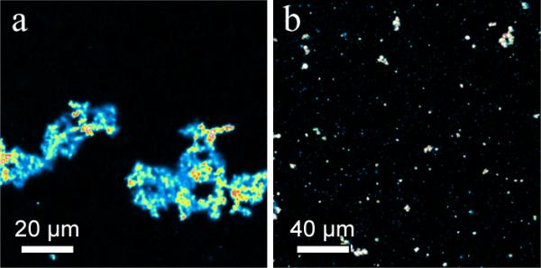
Fig. 4.

(a) Fluorescence image of aggregation of Texas-red DHPE labeled vesosomes after 30 minutes in human blood at 37 °C. (b) Fluorescence images of similar vesosomes as in (a) except for 4 mol% DPPE-PEG750 added to the outer bilayer shell. Aggregation is substantially reduced.
