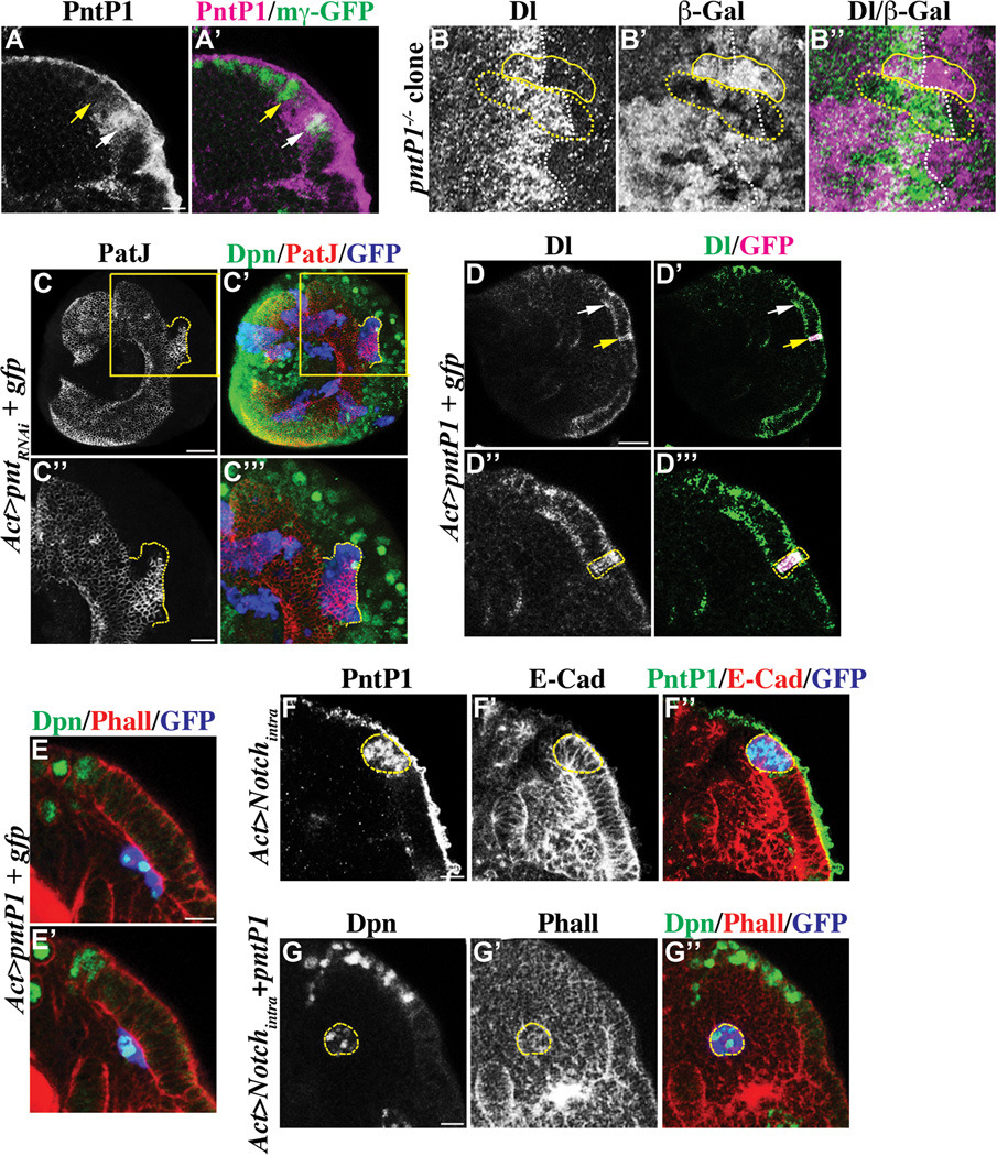
Figure 5. PntP1 promotes conversion of transitioning neuroepithelial cells into immature neuroblasts.
(A–A’) PntP1 becomes transiently expressed in transitioning neuroepithelial cells. The expression of PntP1 largely co-localizes with E(spl)mγ-GFP in transitioning neuroepithelial cells (white arrow), but is undetectable in neuroblasts (yellow arrow). Please note that the antibody against PntP1 shows non-specific background staining on the surface of the brain. The scale bar = 10 µm.
(B–B’’) pntP1 is required for efficient conversion of neuroepithelia into neuroblasts. The conversion of neuroepithelial cells into neuroblasts is significant delayed in the negatively marked pntP1 mutant mosaic clone (outline in dotted yellow line) as indicated by prolonged expression of a high level of Delta. In contrast, neuroepithelial cells in the wild type control twin-spot clone (outlined in solid yellow line) show down-regulation Delta expression (white dotted line) and undergo conversion into neuroblasts synchronously with the surrounding cells outside of the clone.
(C–C’’’) The pnt gene is necessary for conversion of neuroepithelial cells into neuroblasts. Simultaneously reducing the function of pntP1 and pntP2 prevents neuroepithelial cells in the GFP-marked mosaic clone (dotted yellow line) from becoming converted into immature neuroblasts. The scale bar = 20 µm. The higher magnification image of the boxed area is shown in C’’ and C’’’. The scale bar = 10 µm.
(D–E’) pntP1 is sufficient to induce conversion of neuroepithelial cells into neuroblasts. (D-D’’’) Transient over-expression of pntP1 leads to dramatic up-regulation of Delta in neuroepithelial cells in the GFP-marked genetic clone (yellow arrow). The white arrow indicates transitioning neuroepithelial cells. The scale bar = 20 µm. The higher magnification image of neuroepithelia containing the clone (outlined in dotted yellow line) is shown in D’’ and D’’’. (E-E’) Neuroepithelial cells over-expressing pntP1 marked by expression of GFP delaminate inward away from the rest of neuroepithelia, and become converted into neuroblasts prematurely. A superficial optical section is shown in E and a distal optical section is shown in E’. The scale bar = 10 µm.
(F–G’’) Constitutively activated Notch signaling de-sensitizes neuroepithelial cells from PntP1. (F-F’’) Neuroepithelial cells over-expressing Notchintra marked by expression of GFP (outlined in dotted yellow line) accumulate at the medial edge of neuroepithelia. These neuroepithelial cells express PntP1, but do not become converted into neuroblasts. The scale bar = 10 µm. (G-G’’) Expression of PntP1 triggers neuroepithelial cells over-expressing Notchintra marked by GFP (outlined in yellow) to undergo conversion into neuroblasts. The scale bar = 10 µm.

