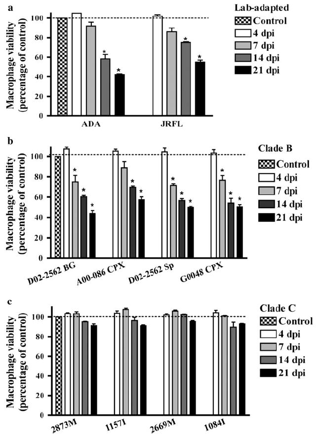
Fig. 2.

Cellular viability of macrophages infected by clade B or C isolates. MDM cultures were infected with 400 TCID50/ml of HIV-1 laboratory-adapted strains (a), clade B (b), or clade C (c) isolates. Supernatants were collected at 4, 7, 14, and 21 days postinfection (dpi). Cell viability was determined by MTT reduction assay and results correspond to the percentage of viable cells normalized to day-matched uninfected control MDM. Values represent SEM from three independent experiments and * denotes p < 0.01 in comparison to control
