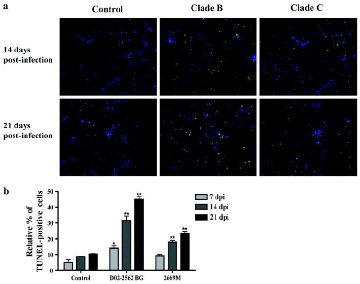
Fig. 6.

Apoptosis of human neurons following treatment with conditioned medium from infected cultures. Human neurons were treated for 3 days with MCM collected from cultures infected for 7, 14, and 21 days with 200 TCID50/ml of D02-2562 (clade B primary isolate) or 800 TCID50/ml 2669M (clade C primary isolate). Apoptotic cells were labeled with TUNEL, and nuclei of total cells were stained with DAPI (a). Photographs have a magnification of ×200. Neuronal apoptosis is expressed as a percentage of TUNEL-positive cells over the total amount of neurons (nuclei staining by DAPI, b). Data values represent SEM and * denotes p < 0.01 in comparison to control
