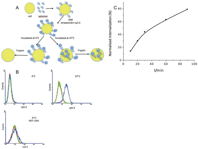
Figure 2. Internalization study showed that KMF2-1a can be internalized specifically into target MCF-10AT1 cells.
A: Schematic of internalization assay. MCF-10AT1 cells were split into 6-well plate. After binding with KMF2-1a-Biotin at 4°C, streptavidin-cy5.5 dye was added. After washing away all the free aptamers and dye and adding medium, one group was incubated at 4°C for 2h and another group was incubated at 37°C for 2h. Trypsin was used to detach and treat cells. B: Internalization of KMF2-1a aptamer. Red line: MCF-10AT1 cells. Green line: library aptamers (negative control) were not internalized. Blue line: Trypsin was applied to remove the membrane-bound aptamers; fluorescence shift existed with KMF2-1a incubated cells at 37°C, but not at 4°C. However, KMF2-1a could not bind to negative control cell MCF-10A1 and could not be internalized by MCF-10A1 cells. C: Quantitative analysis of KMF2-1a internalization with time. The fluorescence intensities of cells that had internalized aptamers were normalized to those of cells at 4°C. Flow cytometry data showed increasing fluorescence intensity inside target cells, indicating increasing internalization of aptamers.

