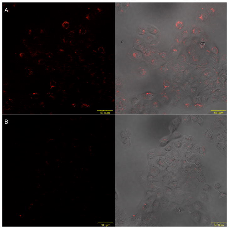
Figure 3. Internalization study of KMF2-1a-TAMRA into MCF-10AT1 cells by confocal microscopy.
(Left: fluorescence signal; Right: overlay of transmitted and fluorescence signal.) A: fluorescence signals were seen from inside the cells after incubation of KMF2-1a-TAMRA with cells for 2 hours at 37°C. B: Lib-TAMRA could not be internalized into cells at 37°C, even after an incubation of two hours.

